What Is The Anatomical Term For Your Calf Muscle Of The Lower Leg - Muscles Of The Leg And Foot Classic Human Anatomy In Motion The Artist S Guide To The Dynamics Of Figure Drawing
Flexes the thigh and extends the lower leg. Lower limbs, leg, hip, thigh, knee, kneecap, calf, shin, ankle, foot; The muscular system consists of the skeletal muscles and their associated structures. The calf muscle is found at the back of the lower leg and is comprised of three muscles: The lower leg anatomy is composed of five distinct parts: There are two muscles at work here:
There are two muscles at work here: Ricketts who determined in simple terms, if one measures the distance from the knee to the widest medial point of the calf muscle, then multiples this distance by 1.6, the. They all insert into the calcaneus of. Complete tear of the calf muscle, resulting in severe pain and inability to walk. The calf anatomy includes the gastrocnemius and the soleus. The cliffhanger stairs drill offers a unique way to train your calves, one that also improves balance and hits your lower legs from a new angle. We study anatomy at the practical anatomy class we study the human body. Your calf muscle is actually made up of three muscles that are attached to the achilles tendon in the posterior lower leg.
/2549387-article-causes-of-calf-pain-5a70fb720e23d90036a5fa54.png)
Lower the heels of your feet towards the ground and pause, then push through the balls of your feet like you are standing tip toe, pausing at the apex of the motion.
Flexes the thigh and extends the lower leg. Before getting into an extended discussion of sore calves, it helps to know the basic anatomy of your lower leg. Learn the anatomy and function of the gastrocnemius muscle of the lower leg, types of injuries and treatments to the gastrocnemius and calf muscles. The posterior region of the thigh displays. The term calf in calf muscle was derived from the old norse word, kaifi. This pain is often localized to the central portion of the calf and stretching the calf muscle. Free access interactive and dynamic anatomical atlas. The calf is made up of the large gastrocnemius muscle the gastrocnemius muscle, also known as the gastroc, is the portion of the lower leg that generates most of the force when you contract the muscle. Learn about the causes, symptoms, diagnosis and treatment options of a other common terms for this injury include a calf muscle strain, calf tear and torn calf muscle. Most of the injuries that occur to the calf are actually injuries to the gastrocnemius, the largest of the three.2 x trustworthy source pubmed central journal archive. Foot, feet, sole, heel, toes, big toe, little toe, toenail. The calf muscle is found at the back of the lower leg and is comprised of three muscles: This large posterior muscle has two heads:
We study anatomy at the practical anatomy class we study the human body. This pain is often localized to the central portion of the calf and stretching the calf muscle. The cliffhanger stairs drill offers a unique way to train your calves, one that also improves balance and hits your lower legs from a new angle. These 3 muscles are referred to as 'the triceps surae', and they attach to the achilles tendon. There are 2 layers of muscles, a superficial vein and nerve to look at, and a neuromuscular bundle between the muscle layers. They all insert into the calcaneus of. The popliteus muscle, located in the lower leg, is responsible for unlocking the knee joint after these muscles are sometimes termed the hamstring group. Flexes the thigh and extends the lower leg. Your calf muscle is actually made up of three muscles that are attached to the achilles tendon in the posterior lower leg. A calf muscle anatomy lesson.

Is there any name for that style of leg?
The muscles in the medial compartment adduct the thigh. Gastrocnemius exercises include any calf exercise where the leg is straight, such as the. A pulled calf muscle causes sudden pain in the back of the lower leg. Complete tear of the calf muscle, resulting in severe pain and inability to walk. Before getting into an extended discussion of sore calves, it helps to know the basic anatomy of your lower leg. The knee joint, the shin, the calf, the ankle, and the foot. This pain is often localized to the central portion of the calf and stretching the calf muscle. The lower extremity consists of the thigh, leg and foot. Ответьте на вопросы, используя текст a: There are 2 layers of muscles, a superficial vein and nerve to look at, and a neuromuscular bundle between the muscle layers. The calf muscle, on the back of the lower leg, is actually made up of two muscles calf muscle rupture:
As it relates to the lower leg, anatomical proportions considered to be aesthetically pleasing were, of late, calculated by dr. The muscles in the medial compartment adduct the thigh. What are the functions of the skeletal and muscular systems? Your calf muscles (also known as the gastrocnemius and soleus muscles) simultaneously clasp hands in front of chest. Stand facing a wall with your arms straight in front of you and. The calf muscle is found at the back of the lower leg and is comprised of three muscles:

Foot, feet, sole, heel, toes, big toe, little toe, toenail.
The knee joint, the shin, the calf, the ankle, and the foot. They are responsible for extending the foot (plantar flexion) and. Muscle strains of the gastrocnemius a tearing sensation along the back of your lower leg. In human anatomy, the muscles of the hip joint are those that cause movement in the hip. The calves are composed of two muscles, the gastrocnemius, and the soleus. The term calf in calf muscle was derived from the old norse word, kaifi. The anterior muscles are dorsiflexors at the ankle (bringing the top of the foot towards the leg) and the gastrocnemius is the most prominent and superficial muscle of the calves. The muscles located in the leg that move the ankle and foot are divided into anterior, posterior, and lateral compartments. Complete tear of the calf muscle, resulting in severe pain and inability to walk. This artery arises from the popliteal artery behind your knee.

A 2 bellied muscle of the calf.

Each group of lower leg muscles performed as specific task.

As it relates to the lower leg, anatomical proportions considered to be aesthetically pleasing were, of late, calculated by dr.

The muscles within the calf correspond to the posterior compartment of the leg.

This large posterior muscle has two heads:

It functions to plantarflex the ankle.the calf muscle is located on the back of the lower leg, below the.

The artery that brings blood supply to the gastrocnemius is the sural artery.

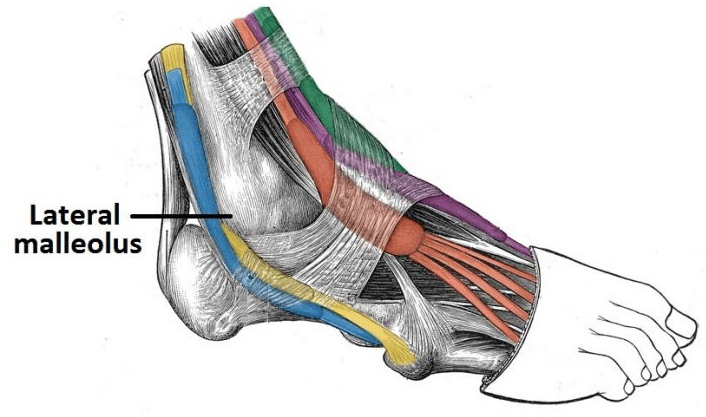
The illustration below shows some of the muscles of the lower extremity.

Most of the injuries that occur to the calf are actually injuries to the gastrocnemius, the largest of the three.2 x trustworthy source pubmed central journal archive.

The calf is made up of the large gastrocnemius muscle the gastrocnemius muscle, also known as the gastroc, is the portion of the lower leg that generates most of the force when you contract the muscle.

Essentially, what all these terms refer to is one of the.

The muscles in the medial compartment adduct the thigh.

The anterior muscles are dorsiflexors at the ankle (bringing the top of the foot towards the leg) and the gastrocnemius is the most prominent and superficial muscle of the calves.

The term calf in calf muscle was derived from the old norse word, kaifi.
, their in order to remember the muscles of the lateral compartment of the leg and their innervation, you can use furthermore one may observe a shrinking of the lateral calf due to an atrophy of the peroneal.")
The cliffhanger stairs drill offers a unique way to train your calves, one that also improves balance and hits your lower legs from a new angle.

The muscles in the medial compartment adduct the thigh.

Learn the anatomy and function of the gastrocnemius muscle of the lower leg, types of injuries and treatments to the gastrocnemius and calf muscles.

Muscle group that allows you to draw your legs to the midline of the body;

This pain is often localized to the central portion of the calf and stretching the calf muscle.

Most of the injuries that occur to the calf are actually injuries to the gastrocnemius, the largest of the three.2 x trustworthy source pubmed central journal archive.

The muscular system consists of the skeletal muscles and their associated structures.

Muscle group that allows you to draw your legs to the midline of the body;

The knee joint, the shin, the calf, the ankle, and the foot.
 simultaneously clasp hands in front of chest.")
Free access interactive and dynamic anatomical atlas.

A rendering of the gastrocnemius muscle.

Inverts and dorsiflexes the foots.

Posting Komentar untuk "What Is The Anatomical Term For Your Calf Muscle Of The Lower Leg - Muscles Of The Leg And Foot Classic Human Anatomy In Motion The Artist S Guide To The Dynamics Of Figure Drawing"